


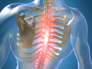
Unnatural changes in spinal cartilage and bones can lead to the development of the disease. According to the ICD-10 code, the disease refers to the location of M42 and is called thoracic osteochondrosis. The pressure on the middle of the spine is less than that of the lumbar and cervical spine, but the deformity is difficult to heal. Because the sternum is round and the load is unevenly distributed, osteophytes and other dysplasia appear.
Symptoms and signs
The disease occurs in the nucleus pulposus of the intervertebral disc and spreads to the vertebral segments such as the annulus fibrosus, ensuring the mobility of the spine. The changes are manifested as compression, reflexes, or mixed neurological disorders and syndromes.
Pain is manifested as physical exertion. There are different types of feelings:
- Mild persistent pain in the chest area is called back pain;
- Sharp and sharp colic, causing difficulty in inhaling or exhaling, causing muscle immobility-the back.
The symptoms and treatment of thoracic osteochondrosis depend on the degree of wear and tear of bone organs and the stage of aging, which can be systemic and local.
Symptoms include:
- Damage to the peripheral process of nerves (neuralgia), which is characterized by painful episodes along the intercostal vasoconstrictors;
- Concentration of pain on the left side of the chest or strong pain of shingles;
- Decreased spine mobility in the chest area;
- Numbness in arms and hands;
- Decline in sexual function;
- Pain in the visceral area can cause heart, stomach, and liver;
- Back pain in the neck, cheekbones and head, cough or lump in the throat;
- Arrhythmia, tachycardia, fever.
The signs of osteochondrosis are disguised as manifestations of related diseases, so the symptoms are not clear. Spinal nerves are concentrated around the spine; when they are clamped, signals are sent to different parts and organs of the body.
Causes of osteochondrosis
There is no exact information about which factors deform the intervertebral disc. A common cause of osteochondrosis is scoliosis or curvature, which is more common in childhood and adolescence.
The theory considers these factors of vertebral deformity:
- Dysplasia;
- Hormones
- Blood vessel;
- Functional
- Inside roll
- Infectious
- immunity;
- Metabolic disorders;
- Mechanical;
- Genetic.
Degeneration and aging of bones and cartilage occur as a result of previous exposure to adverse conditions. Atrophic degeneration of the spine is predetermined by genetic factors, and diseases with clinical symptoms appear under the influence of exogenous and endogenous environments.
When the destruction process of complex substances outweighs their synthesis, consequences in the form of complications of vertebral work occur. Deterioration occurs when the disk power is disturbed and useful elements are scarce. The penetration of elements and alienation products decreases, cell viability decreases, and some cells accumulate due to self-destruction. The production of complex proteins is reduced, and collagen fibers are destroyed.
The mechanical effect on the annular connective tissue is increased, the layered structure is disordered, and the fiber skeleton is torn. The intervertebral disc bruises under the influence of biomechanical factors and body movement, and its fixation ability is reduced. Due to the decrease in hydrostatic pressure, blood vessels and nerves can grow into the valve annulus.
diagnosis method
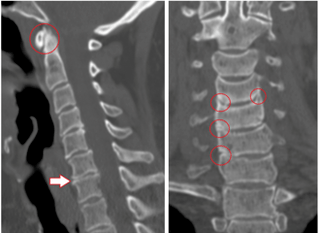
In the identification process, identify nerve roots, pain, reflexes, rigidity, autonomic nerves and vascular factors. The best inspection method is difficult to determine, because in each case, the diagnosis is performed separately.
The main methods are:
- X-ray diagnosis;
- CT scan;
- Magnetic resonance imaging.
X-ray analysis of the condition of the spine, the image is given in oblique projection, lateral projection and direct projection. Sometimes, for a photo, a person will bend, straighten, or bend to one side.
Contrast agents are divided into the following studies:
- Poliomyelitis-inject 20 to 40 milliliters of air into the spinal canal;
- Angiography-Inject 10 ml of contrast medium into the spinal canal and take 7 to 9 images within 2-3 seconds;
- Myelography-inject colored liquid into the subarachnoid space, and then transilluminate the structure;
- Intervertebral discography-the dye is injected directly into the intervertebral disc for local inspection.
Computed tomography scans to evaluate the bone and tissue structure, and the state of blood vessels. The painless method can capture 3D images in a few minutes.
Benefits of CT:
- Fast detection speed;
- Screen "dumb" areas during exercise diagnosis;
- The possibility of multiple spiral angiography;
- Recognize long objects by obtaining high-quality low-thickness cuts.
MRI uses the magnetic field of a machine to generate hydrogen atoms in the human body in parallel with the movement. The particles send out a signal and record the response. The tomograph recognizes the waves and displays the results on the screen. Using MRI, there is no radiation, this method is less dangerous, but it is not recommended for pregnant women.
Treatment and prevention
It is necessary to treat osteochondrosis in several stages, the complexity of which depends on the severity of the disease, contraindications and physical resources.
method:
- Drugs and drugs;
- Physiotherapy methods, exercises to remove clips, alleviate the patient's condition;
- Operation.
There is a direction of exercise therapy in which rehabilitative gymnastics can be used to treat spine problems in the form of hernias and spondylopathy. In addition, a method of recovery after surgery has also been developed.
Yoga practice helps adult men, women, and children overcome pain. The warning is mainly about psychological attitudes.
drug
The medication is prescribed by a neurosurgeon or neuropathologist based on the card and medical history. The patient takes medicine in the hospital or at home, mainly to follow the doctor's advice, not to deviate from the taking plan.
Commonly used drugs:
- Non-steroidal anti-inflammatory drugs can relieve pain, fever and inflammation;
- Muscle relaxants reduce the muscle tone of the bones;
- Hormones reduce neuralgia;
- Vitamins B2, B6, B12, A and C are taken during remission and for simple prevention;
- Diuretics can relieve swelling and release compressed root nerves;
- Neurometabolic stimulants improve the metabolism of nerve tissue;
- The chondroprotectant can restore the cartilage of the vertebrae after damage.
Sometimes, patients do not take medication during the first stage of the onset of unpleasant sensations. Exercise is enough, use a massager.
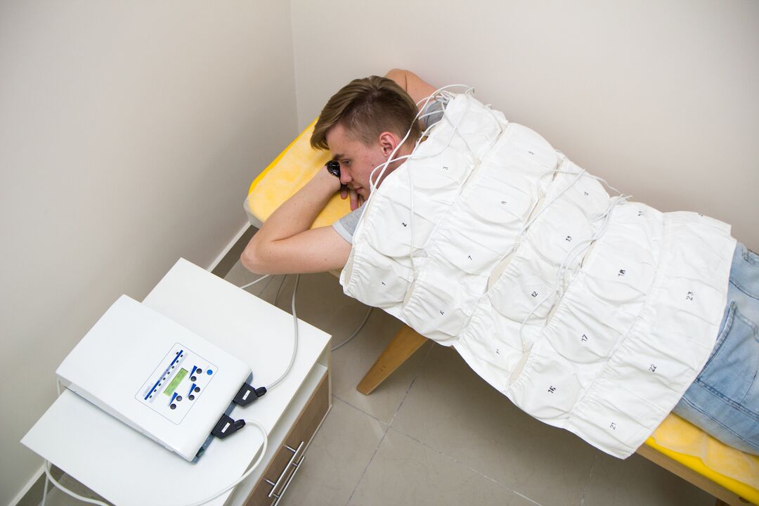
physiotherapy
This exposure is used in combination with drug therapy or alone. In addition, rest in bed and apply heat to the affected area. Folk recipes are used to relieve pain.
Physical therapy in medical institutions includes the following procedures:
- Ultrasound and sonic introduction;
- Shock wave therapy
- Stabilizer impact;
- Laser Treatment;
- electrotherapy;
- Magnetic wave
- Mud therapy and balneotherapy;
- massage.
Ultrasound involves the impact of high-frequency waves on tissues, thereby reducing pain sensitivity. Through ultrasound introduction, analgesics and anti-inflammatory drugs are added to better deliver the drugs to the affected area.
Shockwave therapy is to transmit sound waves to the painful area to improve blood circulation and accelerate metabolism. Detensor therapy involves using the patient's body weight to stretch the spine.
Laser therapy is based on the laser generated by helium-neon to activate the bioelectric current in nerve fibers. The laser acts on the inflamed nerve roots in the paravertebral area along the thoracic spine.
Electrotherapy improves the nutrition and metabolism of the products in the tissue, and the pulse current affects the nerve sensory terminals. Low-frequency waves can relieve acute pain and be used as an initial aid.
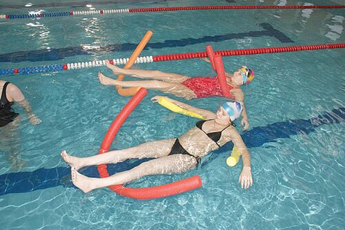
Magnetic therapy is used to relieve swelling, cramps and inflammation. The magnetic wave sensor is placed on the affected chest area. Balneotherapy and mud therapy include swimming in a swimming pool, bathing, and contrast showering for treatment and recovery. Metabolism returns to normal, blood flow to the affected area is accelerated, pain and inflammation are reduced.
The therapeutic massage for thoracic osteochondrosis is vacuum, acupuncture points and lymphatic drainage, which can improve blood microcirculation, tissue nutrition and strengthen muscles. These courses are conducted by qualified experts, and if you believe in the spine of an amateur, dangerous consequences may occur. Massage is prescribed after the acute phase, and the first time should not exceed 10 minutes.
operation treatment
If medical treatment, massage, and other procedures do not relieve the condition, the patient is advised to undergo surgery.
The intervention is divided into 2 stages:
- Eliminate the cause of severe pain (decompensation);
- The stability of the spine.
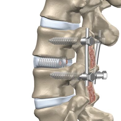
Through the posterior approach, a facial incision is performed because the small joints can compress the nerves. Foraminal incision is the expansion of a nerve root canal through which the nerve leaves the vertebra. Laminectomy removes the back of the vertebrae, which protect the spinal canal and compress the brain due to deformities. Laminectomy involves expanding the opening of the spinal canal where the spinal cord is located while removing individual fragments in the posterior area of the vertebrae.
If there is a herniation (protrusion of the intervertebral disc into the spinal canal cavity) or hernia herniation into the spinal canal, anterior surgery is performed.
The methods used in the previous decompression are as follows:
- Discectomy-removal of the entire intervertebral disc or a part of it;
- Vertebrectomy-the entire vertebrae and adjacent intervertebral discs are removed and then implanted.
Discectomy and vertebral body resection cause column instability and increase the risk of neurological defects. Use rigid fixation or fusion (fusion) of three vertebrae.
Prevent chest osteochondrosis
The deterioration of the disease will reduce the work ability and the quality of human life, so special attention should be paid to prevention. As a result, vertebral degeneration appears later and disability is avoided.
Ways to prevent diseases:
- Reduced physical activity on the spine;
- If you don’t change the outriggers, you can’t stand for a long time, you can lean on a temporary built object or wall;
- It is not recommended to sit at the desk for a long time, and when using the computer to work, you need to take active rest and move around;
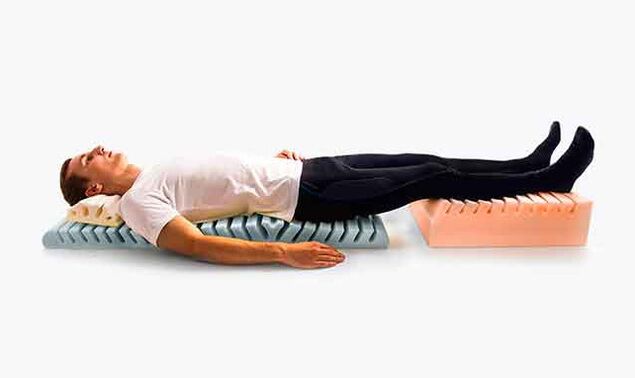
- Choose mattresses and orthopedic headrests for sleep;
- When running and walking, avoid sudden turns and jumps, and walk in shoes with small heels;
- Load no more than 10 kg, gradually lift from the sitting position.
In the car, backrests and headrests need to use pillows, and the driver's seat must be rigid. Work cannot be done in a semi-tilted posture, you can stand or sit. Developed muscles support the bones, so they focus on feasible physical exercises and exercises.
Possible complications
The disease takes a long time to develop, and sometimes the painful symptoms do not appear immediately. Any degenerative changes in the thoracic area will lead to the appearance of pathology.
Types of complications:
- Cardiovascular pathology followed by myocardial infarction or angina pectoris;
- Chest pain caused by intercostal neuralgia or peripheral nerve inflammation with root compression;
- Herniated disc.
Complications of advanced osteochondrosis will occur, so early and timely treatment will help avoid accompanying diseases.