


Hip Joint Hip Joint Disease (HJ) is a degenerative dystrophic disease that affects cartilage and bone tissue. In medical articles, it can have different names: deformable hip joint disease, hip DOA, osteoarthritis. All these terms refer to the same pathology-arthropathy, but "hip arthropathy" is a narrower concept that characterizes the failure of the hip joint.
Cartilage is the first part of joint disease, then bones and surrounding structures-ligaments and muscles-are involved in the pathological process. If the bones change, add the prefix "osteo" after the word "arthropathy". In advanced cases, the joints are deformed and they are already talking about deformed joints (osteoarthritis).
General characteristics
Deformable osteoarthritis of the hip is the second most common disease after knee arthritis. Due to the deep position of the hip joint, bone deformities can be undetected for a long time, and only the X-ray images taken later will show changes.
The development of this disease is affected by many factors, including an inactive lifestyle, trauma, and metabolic disorders. It is precisely because of the particularity of modern life that there is often no room for physical education, arthropathy affects more and more people. In addition, the peak incidence falls in the middle-aged group-from 40 to 60 years old.
refer to:Hip joint disease often affects women more than men.
Development mechanism
The thigh joint is made up of two bones: the femur and the ilium (pelvis). The femoral head enters the acetabulum of the pelvis and remains motionless during exercise-walking, running. At the same time, the articular surface of the femur can move in multiple directions, providing flexion, extension, abduction, adduction and rotation (rotation) of the thigh.
During physical activity, the femur moves freely in the acetabulum due to the cartilage tissue covering the joint surface. Hyaline cartilage is known for its strength, firmness and elasticity; it acts as a shock absorber and participates in the load distribution during human movement.
Inside the joint is synovial fluid-the synovial membrane-which is essential for lubricating and nourishing cartilage. The entire joint is enclosed in a dense thin capsule surrounded by powerful muscles of the thigh and buttocks. These muscles also act as shock absorbers to prevent hip joint injuries.
The development of hip joint disease begins with changes in joint fluid, which becomes more viscous. Due to lack of water, the cartilage does not get enough nutrients, begins to dry, loses smoothness, and cracks appear.
The bones can no longer move freely as before and rub against each other, which can cause micro-damage to the cartilage. The pressure between the bones increases and the cartilage layer becomes thinner. Under the influence of increased pressure, bones gradually deform and local metabolic processes are disrupted. In the later stages, the leg muscles are significantly atrophy.
reason
Deformed joints of the hip joint can be primary and secondary. It is not always possible to determine the cause of primary joint disease. Secondary arthropathy appears in the context of existing diseases, namely:
- Congenital dislocation of the hip or hip dysplasia;
- Perthes disease (aseptic necrosis of the femoral head);
- Hip arthritis of infectious, rheumatic or other origin;
- Pelvic bone injury-dislocation, fracture.
Hip dysplasia is a congenital deformity that sometimes will not be clinically manifested for a long time, and in the future (25-55 years old) can lead to the development of dysplastic hip arthropathy.
Coxo arthropathy can be left, right and symmetrical. In primary arthropathy, concomitant diseases of the musculoskeletal system are often observed-especially osteochondrosis and knee joint disease.
There are also some risk factors that can lead to the occurrence of diseases:
- Overweight and overload overload the joints;
- Violation of blood circulation and metabolism;
- Hormonal changes;
- Curved spine, flat feet;
- Advanced age
- Decreased physical strength;
- Genetic.
It should be noted that hip joint disease itself is not hereditary. However, certain characteristics of the metabolism or structure of connective tissue can create prerequisites for the development of joint joint disease in children in the future.
Symptoms of hip joint disease
The main symptom of hip joint arthropathy is pain in the hip and groin area, with varying degrees of pain. Also noticed stiffness and stiffness during exercise, decreased muscle volume, shortening of affected limbs, and gait changes due to claudication.
Hip arthropathy usually progresses slowly, causing discomfort at first, and mild pain after exertion. However, over time, soreness will increase and appear at rest.
The typical manifestation of pathology is difficulty in hip abduction, that is, a person cannot "straddle" on a chair. The presence and severity of the signs of hip joint disease depend on its degree, but pain syndrome is always present.
Arthropathy of the hip is divided into three degrees or types, which vary according to the severity of the injury and the accompanying symptoms:
- 1 degree. The thigh is not always painful, but periodic pain, mainly after walking or standing for a long time. The pain syndrome is confined to the joint area, but sometimes spreads from the legs to the knees. 1st-degree hip joint disease does not atrophy the muscles, does not change the gait, and fully retains the athletic ability;
- level 2. Pain will increase, not only after running or walking, but also when resting. The pain is more often concentrated in the thigh area, but it can spread down to the knee. Under heavy load, it hurts to step on the injured limb, so the patient begins to protect the leg and limp. The range of joint motion is reduced, and it is particularly difficult to move the leg to one side or rotate the hip;
- 3 degrees. The soreness will become permanent and will not subside even at night. Gait is obviously impaired, independent movement is obviously complicated, and the patient is on crutches. The range of motion is obviously limited, and the hip muscles and the entire leg muscles including the calf atrophy.
- Due to muscle weakness, the pelvis is tilted forward and the affected limb is shortened. In order to compensate for the difference in the length of the limbs, the patient tilts the body to the affected side while walking. This can cause the center of gravity to shift and increase the pressure on the affected joints.
Osteoarthritis or osteoarthritis?
Arthritis is an inflammation of the joints. It can be an independent disease or it can develop in the context of a systemic disease (such as rheumatism). In addition to the inflammatory response, the symptoms of osteoarthritis (especially in the advanced stages) also include restricted mobility and changes in joint shape.
The core of the changes in joint degenerative dystrophy is the destruction of cartilage tissue, which usually leads to inflammation. This is why arthropathy is sometimes called arthropathy-arthritis. Since arthropathy is almost always associated with joint deformation, the term "osteoarthritis" applies to it.
refer to:According to the International Classification of Diseases (ICD-10), osteoarthritis and osteoarthritis are variants of the same pathology.
Diagnosis of hip joint disease
The diagnosis of "hip joint hip joint disease" is made based on examinations, patient complaints and examination results. The most informative method is X-ray: in the picture, you can see the extent of joint damage and the cause of the disease.
For example, in hip dysplasia, the acetabulum is flat and inclined, and the cervical diaphyseal angle (the inclination of the femoral neck in the vertical plane) is greater than normal. The deformity of the part of the femur near the joint is characteristic of Perthes disease.
3rd degree hip arthropathy is characterized by narrowing of the joint space, expansion of the femoral head, and multiple bone growths (osteophytes).
If the patient fractures or dislocates, signs of trauma can also be seen on X-rays. If a detailed assessment of the condition of bones and soft tissues is required, magnetic resonance imaging or computed tomography can be prescribed.
Differential diagnosis of the following diseases:
- Knee joint disease;
- Osteochondrosis and nerve root syndrome appearing in its background;
- Trochanteritis (inflammation of the trochanteric bone of the thigh);
- Ankylosing spondylitis;
- Reactive arthritis.
The reduction in muscle volume that accompanies 2nd and 3rd degree hip joint disease can cause pain in the knee joint area. In addition, knee pain is often more painful than the hip joint itself. To confirm the diagnosis and rule out knee joint disease, X-rays are usually sufficient.
Spinal diseases-osteochondrosis and nerve root compression-the pain is very similar to hip joint disease. However, it happened unexpectedly, after an unsuccessful exercise, a sharp turn of the body, or a weightlifting. The pain starts in the hip area and spreads down to the back of the leg.
Nerve root syndrome is characterized by severe pain when lifting straight limbs from a supine position. However, there is no difficulty in the process of abducting the leg to one side, just like hip joint disease. It is worth noting that osteochondrosis and arthropathy of the hip are often diagnosed at the same time, so a comprehensive examination is required.
Rotoritis or trochanteric bursitis develops rapidly. In contrast to arthropathy, arthropathy can progress slowly over several years or even decades. Pain syndrome can develop within a week or two, but it is very severe. The cause of trochanteritis is trauma or excessive exercise. Movement is not restricted and the legs are not shortened.
Ankylosing spondylitis and reactive arthritis may also be accompanied by symptoms similar to hip joint disease. The hallmark of this type of disease is pain mainly at night. The hip may be very painful, but when walking and moving, the pain will subside. In the morning, the patient is worried that the stiffness will disappear after a few hours.
Treatment of hip joint disease
Hip joint disease can be treated conservatively or surgically. The choice of treatment depends on the stage and nature of the pathological process. If a degree 1 or 2 disease is diagnosed, it is treated with medication and physical therapy. After alleviating the acute symptoms, therapeutic exercises and massage were added. If necessary, prescribe special diets.
The earlier the hip joint disease is detected and treated, the better the prognosis. With the help of drugs and treatments, you can significantly slow down the pathological process and improve the quality of life.
Non-steroidal anti-inflammatory drugs (NSAIDs) are used to relieve pain and inflammation. It should be noted that anesthesia is performed in the shortest possible process, because NSAID drugs will negatively affect the digestive tract and slow down the regeneration process of cartilage tissue.
With the help of chondroprotective agents, the recovery of cartilage can be accelerated. However, these funds are only effective in the early stages of the disease, when the hyaline cartilage is not completely destroyed. Chondroprotective agents are prescribed in the form of tablets or intra-articular injections.
In order to improve the blood supply to the joints, vasodilators are used. For muscle cramps, muscle relaxants are recommended.
For persistent pain syndromes that are difficult to eliminate with pills, injections can be made into the hip joint. Corticosteroids can relieve inflammation and pain very well.
Medication can also be supplemented by topical medications-ointments and gels. They have no obvious effect, but they help cope with muscle cramps and relieve soreness.
Physical therapy helps to improve blood circulation and cartilage nutrition. For hip joint disease, procedures such as shock wave therapy (SWT), magnetic therapy, infrared laser, ultrasound, and hydrogen sulfide bath have proven to be very good.
Operation
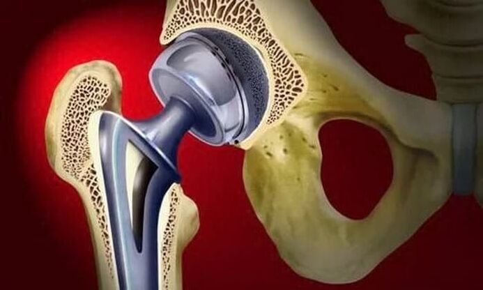
The treatment of stage 3 osteoarthritis can only be done by surgery, because the joints are almost completely destroyed. In order to restore the function of the hip joint, a partial or total joint replacement is performed.
When conservative treatment fails, turn to surgical treatment in advanced cases of arthropathy.
In some prostheses, only the femoral head is replaced by artificial prostheses. A full prosthesis means replacing the femoral head and acetabulum. The operation is performed under general anesthesia, and the majority of cases (about 95%) have fully recovered hip function.
During the recovery period, antibiotics are prescribed to patients to prevent infection complications. The stitches were removed on days 10-12 and exercise therapy started. The attending physician helps the patient learn to walk and correctly distribute the load on the surgical limb. Exercise is an important step to increase muscle strength, endurance and elasticity.
It takes an average of 2-3 months after surgery to regain the ability to work, but for the elderly, this time can be as long as 6 months. After the rehabilitation is completed, the patient can fully move, work, and even exercise. The life of the prosthesis is at least 15 years. In order to replace the worn prosthesis, a second operation is required.
Effect
Without timely and adequate treatment, hip joint disease will not only significantly worsen the quality of life, but also lead to disability and disability. Already in the second stage of joint disease, the patient was assigned to the third disability group.
When the affected limb is shortened by 7 cm or more, when a person moves only with the help of temporary means, the second group will be assigned. The first group of disability was patients with third-degree hip joint disease, accompanied by complete loss of exercise ability.
The indications for the medical and social examination (MSK) are:
- The course of joint disease is long, more than three years, and it deteriorates regularly. The frequency of attacks is at least 3 times every 12 months;
- Received prosthesis surgery;
- Serious impairment of limb musculoskeletal function.
prevention
The main measures to prevent hip joint disease are diet (if you are overweight) and regular but moderate physical activity. It is very important to avoid injury and hypothermia in the pelvic area.
In the presence of risk factors for joint disease, as well as all patients with confirmed diseases, swimming is beneficial. Sports such as running, jumping, football and tennis are not recommended.