


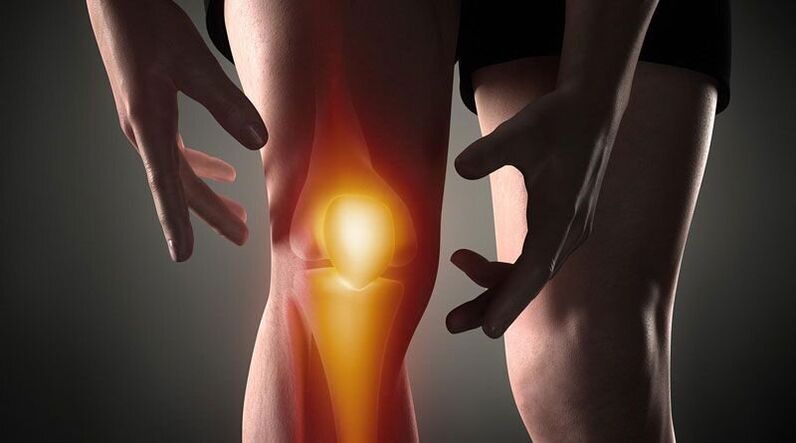
Knee pain-This is a sign of a pathological process affecting the cartilage, bone or soft tissue structure of the femur-tibia and femur-patella joints. Joint pain can be based on trauma, inflammation, and degenerative diseases of joint organs and structures around joints. Patients may complain of severe, painful, burning, throbbing, and other types of pain during rest or when moving, supporting, bending, and stretching the leg at the knee. Pathological diagnosis of etiology includes instrument imaging methods (Rg, ultrasound, CT or MRI, arthroscopy), joint capsule puncture, biochemical and immunological analysis. Before the diagnosis, rest, joint fixation, non-steroidal anti-inflammatory drugs and analgesics are recommended.
Causes of knee pain
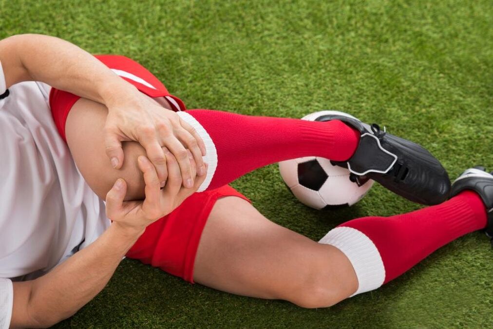
trauma
They are usually the result of family trauma and are common in athletes: runners, jumpers, sports participants. Caused by a fall, direct impact, or twisting of the leg. Manifested as severe pain at the time of injury. In the future, the pain syndrome will become less obvious and will be accompanied by increased edema. Scratches and bruises may appear. As the frequency increases, the following injuries will be found:
- Knee injury. . . Occurs when you fall on your knee or hit your knee directly. At first, the pain is intense, burning, sometimes burning, but it can be tolerated, and later-dull pain, pain, and aggravated by exercise. Bruising is possible. Leg support is retained. Sometimes knee joint injury will be accompanied by hemorrhage in the joints. In this case, the joints gradually increase in volume and become spherical, and the pain syndrome will increase the feeling of pressure or bursting.
- Broken ligament.It was discovered after twisting the leg, and it was forced to twist, bend, or overextend in a non-physiological position. The sensation of pain is stronger than a bruise; while the pain occurs, a person can feel how something is torn (similar to how ordinary tissues are torn). It is accompanied by significant restrictions on movement, support, and limb twisting, and hemorrhage in the joints increases rapidly.
- Intra-articular fracture. . . They are detected by impacts, drops and twists of the legs. In the event of an injury, a person feels very sharp, often intolerable severe pain, and sometimes a crunching sound is heard. Patients with intra-articular fractures describe their own feelings like this: "The pain is so dark that the eyes are so dark that the world no longer exists, and you don't understand anything. " Then, the pain became less severe, but the intensity was still high. Support is usually impossible, and movement is almost completely restricted. Edema and hemorrhage progressed rapidly.
- dislocation.It is the result of a blow or a knee fall. Dislocation of the patella will cause severe pain, accompanied by a sensation of bending of the calf and displacement of the knee. It cannot be moved, and the reference function can be saved. On the front surface of the knee, a significant deformation can be seen, which then becomes smooth due to increased edema. Sometimes hemorrhage will join.
- Pathological fracture.They develop with minor injuries and are the result of decreased bone strength in osteoporosis, osteomyelitis, tuberculosis, and bone tumors. Pain is a sore, dull pain, a pain syndrome reminiscent of bruising. Signs of pathological fractures include restricted or unsupported legs, unstable knees, sometimes deformities, and bone tightening during exercise.
- The meniscus is damaged.Meniscus tears are formed when twisting, impacting, strongly forcing the knee to bend or stretch, and making sharp turns with a fixed leg. At first, a person will feel a peculiar clicking sound and intense shooting pain deep in the joints. Then the pain will be reduced, but sometimes it will become diffuse-burning, rupturing, and intensifying when trying to support and move. The volume of the knee increases due to edema and hemorrhage in the joints. Support becomes impossible and movement is severely restricted.
Inflammatory pathology
They can be infectious and non-infectious (post-traumatic, toxic allergy, metabolic, post-vaccination). The sufficient blood supply of the synovium and tissues around the joints promotes the rapid development of inflammation in response to direct and indirect effects, and a large number of nerve endings cause obvious pain responses. The inflammatory process is often accompanied by synovitis (the accumulation of sterile fluid in the joints), and pus may accumulate when infected.
- arthritis.Gonorrhea occurs after injury, sometimes complicating infectious diseases, and is detected in rheumatism. It may be acute or chronic. Knee pain is usually dull pain, soreness, compression, or stretch. At first, the pain is not severe and intermittent, but it gets worse at night or after exercise. Then the beginning pain joins, and the intensity and duration of the pain syndrome increases. The joints are swollen, the skin above them becomes red, and the temperature rises. With synovitis, the contour of the knee becomes smooth and there is a feeling of bursting. With suppuration, the severity of the pain increases sharply, they become convulsive and lack of sleep.
- Synovitis.It is not an independent disease and complicates many acute and chronic pathologies of the joints. It is formed within a few hours or days. Initially, the pain is trivial or non-existent, and satiety prevails. The knee is spherical, with a lot of fluid, and the skin is shiny. Movement is somewhat restricted. After infection, the pain will become obvious, pulsating, twitching, and will be exacerbated by slight movements and touches.
- Bursitis.Inflammation of the joint capsules located in the patella and popliteal fossa usually occurs when the knee is overloaded and repeatedly injured (for example, sustained support of the knee). For bursitis, the pain is local, blunt, and not strong. It appears in a certain position of the limb. After a characteristic load, it will be relieved when the position of the leg changes. Massage the affected area. If the back pocket is affected, you may feel pain when going up and down the stairs. Sometimes a slight local edema is confirmed. With the suppuration of the bursa, the pain becomes severe, convulsions, baking, and is accompanied by hyperemia, edema of the affected area, and symptoms of systemic poisoning.
- Tendinitis.Usually detected in overweight men and athletes, it affects the ligaments of the patella itself. At first, the pain syndrome only occurs during vigorous exercise, then the standard exercise load, and then during daily physical activity or rest. Tendinitis pain is located directly below the knee, dull pain, pulling, and as the disease progresses, sometimes paroxysmal, and in some cases with mild redness and swelling, and increased pressure. Movement is usually complete, with few slight restrictions. Due to its reduced strength, the ligaments may tear or break.
- Fatty arthritis.Hoff's disease affects the layer of fatty tissue under the patella. It is observed that the knee is continuously overloaded or as a result of an old injury. More commonly, it affects athletes and elderly women. A person will complain of dull pain and limited stretch at the same time. As the pathology worsened, pain began at night, and there was a feeling of instability of the knee joint and instability of the leg arch. When you press the side of the patella, you will hear a slight crack or squeak.
Autoimmune process
The cause of this group of diseases is that with the development of aseptic inflammation of the immune complex of synovium and cartilage, antibodies against normal cells of the body are produced, that is, the phenomenon of vasculitis. In most cases, pathologies are chronic, they can easily progress if left untreated, and are usually the cause of disability.
- Rheumatoid Arthritis.Failure is usually bilateral. There is little activity in the autoimmune process, and the pain is weak or moderate, intermittent, pulling, and oppressive, accompanied by morning stiffness. During moderate activity, patients will complain of periodic and prolonged pain, compression, or burst pain of moderate intensity, not only during exercise, but also at rest. There was stiffness for several hours, moderate recurrent synovitis. With the high activity of rheumatoid arthritis, the pain is intense, diffuse, tired, and wavy, increasing in the first few hours of the morning. The stiffness becomes constant, and a large amount of fluid accumulates in the knee, forming a contracture over time.
- Systemic lupus erythematosus.Joint pain is usually symmetrical, but it may affect one joint. They can occur at any stage of the disease; with the recurrence of SLE, they resemble rheumatoid arthritis. With the low activity of the process, the pain is short-term, non-intensive, local, painful, and stretched. In severe cases, the pain syndrome progresses, the pain is wavy, disrupts night sleep, becomes prolonged, diffuse, increases with exercise, and is accompanied by synovitis, edema, and hyperemia.
- Rheumatism.Arthralgia is one of the first manifestations of rheumatic fever. It appears 5-15 days after the acute infection and affects multiple joints at the same time (usually in pairs). The pain is quite short-lived, but very severe. It transfers from one joint to another and is of different nature, ranging from pulling or pressing to burning or pulsation. The knee was swollen, hot, and the skin on the knee turned red. Movement is severely restricted. A few days later, the severity of the pain reduced and the exercise resumed. In some patients, residual effects in the form of moderate or mild dull pain last a long time.
- Reactive arthritis.It occurs more often in 2-4 weeks after intestinal and genitourinary tract infections, usually involving one or two joints of the lower extremities, combined with urethritis and conjunctivitis. The development of reactive arthritis precedes increased urination, urethral pain and burning, tearing, and eye cramps. The knee pain is severe or moderate, persistent, wavy, painful, tugging, twitching, and is accompanied by restricted movement, deterioration of the general condition, fever, severe swelling and redness of the affected area. The pain and signs of inflammation lasted for 3 months to 1 year, and then gradually disappeared.
Degenerative malnutrition process
They develop due to metabolic disorders in the joints and soft tissue structures around the joints. They have a chronic course that lasts for many years. It is often accompanied by the formation of calcifications, cysts and osteophytes, and deformation of the knee joint surface. With the significant damage of the articular surfaces, they cause significant damage to the movement and support functions, becoming the cause of disability, and requiring the installation of endoprostheses.
- Osteoarthritis.It develops without obvious cause or under the background of various injuries and diseases, and mainly occurs in the elderly and middle-aged people. At first, the pain is weak and short-term, usually stretching or soreness. It occurs with prolonged fatigue and disappears at rest, usually accompanied by a crunching sound. The pain syndrome gradually worsened, the knee began to ache "in the weather", and movement was restricted at night. The distinguishing characteristics of knee joint disease are the onset of pain (it won’t be painful until you "dissipate" it), periodic attacks of sharp cuts due to blockade, burning pain, or shooting pain. During exacerbations, synovitis often occurs, in which the pain becomes persistent, oppressive, and ruptured.
- Meniscus disease. . . It is usually found in athletes that their work involves a significant load on the knee joints. It is manifested as deep local pain on the inner side of the knee joint at the level of the joint space, and is more common in the outer half of the knee joint. Pain worsens during exercise and lessens during rest, which may be dull pain, compression, or pulling. As it progresses, there will be severe shooting pain when trying to move. On the anterior and outer sides of the joint where the pain is projected, a small pain can sometimes be felt.
- Tendinopathy. . . the tendons near the knee are affected. The initial manifestation is a short-term localized shallow pain during the peak period of physical activity. Later, with moderate load and light load, pain sensation appeared, limiting daily activities. Pain is stretching or soreness and is directly related to active movement. It is not detected during passive extension and flexion of the knee joint, and is sometimes accompanied by a squeaking or crackling sound. In the lesion area, the most painful part can be explored. Signs of local inflammation (edema, congestion, hyperthermia) are not obvious or non-existent.
- Osteochondrosis.Children and adolescents are more often affected, and the course of the disease can last for several years. It usually starts with mild claudication or intermittent, non-strong dull pain, which worsens when tired, and disappears when resting. As osteochondrosis progresses, the pain becomes intense, persistent, oppressive, burning, or baking, accompanied by severe claudication, restricted mobility, and difficulty resting on the limbs. Then the pain is gradually reduced, supporting the recovery of function.
- Rickets.It is usually diagnosed in older men and is less common in infants. Arthrochondromatosis presents as moderate undulating dull pain, which usually worsens at night and in the morning. Movement is restricted, accompanied by austerity. Sometimes obstructions occur, which are characterized by sudden and severe shooting pain, inability to move, or severe restriction. With the development of synovitis, the pain is explosive, accompanied by increased knee joint volume, soft tissue swelling and local temperature rise.
Tumor and tumor-like formation
Pain syndrome can be caused by cysts, which are benign or malignant tumors that directly affect the joints or the tissues around them. In addition, knee joint pain can be a warning signal for hypertrophic arthropathy and paracancerous polyarthritis (paraneoplastic syndromes characteristic of lung cancer, breast cancer and other tumor processes).
- Baker's cyst.The hernia of the popliteal fossa is prominent. In the initial stage, it manifests as discomfort or slight local pain at the back of the knee. In the context of an increase in Baker's cyst due to compression of nearby nerves, burning or shooting pain, numbness or tingling in the sole area may occur. When trying to bend the knee as much as possible, the symptoms get worse. In the popliteal fossa, elastic, slightly painful tumor-like structures are sometimes felt.
- Benign tumor.Including chondroma, osteochondroma, non-ossifying fibroma and other tumors. They are characterized by a long-term asymptomatic or low-symptomatic course, and they can manifest as vague and intermittent localized non-severe pain. For large tumors, the formation of solids may be felt, and sometimes it may develop into synovitis.
- Malignant tumors.The most common malignant tumors affecting the joint area are synovial sarcoma, osteosarcoma and chondrosarcoma. They show local dull pain and sometimes have a certain circadian rhythm (worse at night). The intensity of the pain increases, they become sharp, cut, burn or twitch, spread along the knee and adjacent tissues, accompanied by deformation, edema, synovitis, saphenous vein dilation, violation of the general condition, and formation of contractures. On palpation, the formation of painful tumor-like formations is confirmed. When this process begins, the pain is unbearable, unbearable, exhausting, deprives you of sleep, and cannot be eliminated by non-narcotic analgesics.
Intrusive manipulation and manipulation
Pain syndrome is caused by damage to the knee joint tissue during invasive surgery. The severity of the pain directly depends on the trauma of the knee joint operation. As pathogenic microorganisms invade the joint area, the pain is caused by inflammatory changes.
- manipulate.The most common operation is puncture. The pain after the puncture is short-term, not severe, quickly subsided, limited to the projection site of the puncture, and usually occurs on the outer surface of the knee. After the biopsy, the pain may twitch at the beginning, and it will become dull and disappear after a few days.
- operate.After arthroscopy, the pain was moderate, at first very severe, then dull, and subsided after a few days or 1-2 weeks. After the joint incision, the pain syndrome is more severe. Due to obvious tissue damage, the pain symptoms can last up to several weeks. Usually, in the first 2 or 3 days after the intervention, the patient is prescribed analgesics, and then the pain weakens and gradually disappears.
Psychosomatic disease
Sometimes under the influence of psychological factors, knee joint pain occurs without an organic basis (traumatic injury, inflammation, destruction, etc. ). People believe that this kind of pain plays a protective role because it helps reduce emotional stress by transforming experiences into physical sensations. A distinctive feature of this kind of pain is that their nature is uncertain, inconsistent, without obvious changes, and a clear connection with physical activity and other objective predisposing factors. Allergic joint pain has been observed in people who are sensitive to changes in atmospheric pressure.
In addition, knee joint pain irradiation may be related to hip joint disease, lumbar osteochondrosis, Perthes disease, fibromyalgia, and sciatic neuropathy. However, due to these pathologies, other localized pain syndromes usually appear. Other risk factors that increase the likelihood of knee injuries and disease include overweight, occupational exercise, vitamin deficiencies, metabolic disorders, and old age. Hypothermia, stress, physical exertion, and eating disorders may be the triggers for worsening chronic pain.
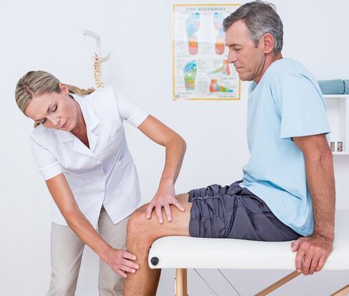
Polls
The diagnostic search algorithm is based on considering the nature of the pain syndrome, its duration, and identifying the accompanying symptoms and events before the onset of knee pain. At the first visit to the doctor (surgeon, surgeon, rheumatologist), visual inspection and palpation of the knee joint is performed to assess the amount of active and passive movement. Taking into account the data obtained, patients can be allocated in the future:
- Laboratory blood test. . . A complete blood count helps to identify hematological changes that are typical of acute infections and inflammatory processes (leukocytosis, elevated ESR), eosinophilia, and allergic reactions. Biochemical and serological research has the greatest amount of information on autoimmune diseases, which is characterized by the formation of specific acute-phase proteins and immunoglobulins (CRP, rheumatoid factor, ASL-O, CEC, DNA antibodies, etc. ).
- Radiography.The basic diagnosis method is that the X-ray of the knee joint is in 2 projections. The existence of pathology is indicated by changes in the contours of the joint head and joint cavity, narrowing of the joint space, changes in the thickness of the endplate, the presence of edge defects at the end of the bone and joint, osteolysis and bone destruction. In certain diseases (meniscal trauma, In Baker’s cyst), contrast arthrography showed the greatest sensitivity.
- Joint ultrasound. . . Knee ultrasound is a fast, cheap, affordable and informative diagnostic method. It allows you to determine whether there is fluid and loose bodies in the joint cavity, and identify the damage and pathological changes (calcification, bleeding, etc. ) of the soft tissues around the joints. They help distinguish the cause of joint pain with high precision.
- CT and MRI. . . They are the preferred method of joint disease of any origin. They are used to assess the nature and extent of pathological changes in more detail to identify typical signs of traumatic, inflammatory and neoplastic lesions in bone structure and soft tissue. CT and MRI of joints usually have limited information content for other instrumental research.
- Joint puncture. . . Execute when there are signs of exudate or accumulation of exudate in the joint capsule. As part of the differential diagnosis of inflammatory, degenerative and tumor diseases, cytology, bacteriology or immunology studies of synovial fluid. In order to determine the diagnosis of knee joint autoimmune injury, tuberculous arthritis, and synovial tumors, it is extremely important to perform a synovial biopsy.
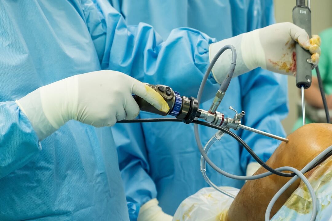
- Arthroscopy. . . The purpose of invasive endoscopic diagnosis can be biopsy sampling to clarify necessary diagnostic information during visual inspection of joint components. In some cases, diagnostic arthroscopy is developed into therapeutic arthroscopy (arthrectomy, meniscus resection, ligament autoplasty, etc. ).
Symptomatic treatment
Taking into account the identified disease, the treatment of the cause of knee joint pain is different. At the same time, symptomatic care is an important part of the comprehensive treatment process aimed at reducing discomfort and improving the quality of life. After injury, it is recommended to apply a cold compress to the knee area immediately-this will help reduce pain sensitivity. Ethyl chloride has a local cooling and anesthetic effect. In all cases, resting your knees will help reduce soreness. It is necessary to limit movement and keep the leg in a position with the least pain. When walking, a fixed bandage is wrapped around the knee, and the limb can be fixed with the help of a plaster model.
In the acute phase of injury or illness, it is strictly forbidden to massage knees, apply heat and wear high heels. The main classes of drugs used for the symptomatic treatment of pain and inflammation are analgesics and non-steroidal anti-inflammatory drugs, which are in the form of ointments, tablets and injections. The listed measures can only temporarily relieve the pain, but cannot eliminate the root cause of joint pain. Therefore, all cases of knee pain require qualified diagnosis and treatment, and some conditions (fractures, dislocations, hemorrhage) require urgent medical care. If the pain is accompanied by changes in the shape of the knee joint (swelling, smoothing of the contour, asymmetry), inability to perform flexion and extension exercises, bounce of the patella, and damage to the knee joint support, the visit cannot be postponed. Limbs.