


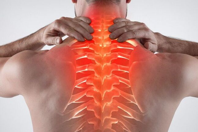
Thoracic osteochondrosis is a common degenerative disease. Thoracic osteochondrosis has specific symptoms, indicating the onset of pathology. In the initial stage, the patient will not feel discomfort, so he is not eager to seek help from a specialist. Over time, the symptoms will intensify, which forces the patient to see a doctor, where a neglected pathology is discovered. You should know what early signs of osteochondrosis can be determined and which treatment is most effective.
What is thoracic osteochondrosis and how does it occur
Thoracic osteochondrosis is characterized by a destructive malnutrition process in the middle of the spine. The damage is located between the 8th and 19th vertebrae. To find the affected vertebrae, it is necessary to conduct accurate diagnostic studies. Thoracic osteochondrosis is usually accompanied by serious complications, including prolapse or hernia. Without complications, this disease is rare, because the destruction of cartilage tissue inevitably leads to the destruction of the entire vertebral framework.
When a patient has circulatory disorders or age-related joint wear, the annulus fibrosus located in the intervertebral disc cavity begins to collapse and lose its normal structure. Due to the slow destruction, micro-cracks will appear in the initial stage, and the nucleus pulposus will seep out from it.
As the internal components ooze out, the fibrous ring begins to weaken, leading to gradual stretching and rupture. When the nucleus pulposus is herniated, intervertebral hernia occurs, which is the most common complication of osteochondrosis. Pathology involves damage to cartilage tissue, which can cause significant discomfort. Severe back pain is also related to neurological syndromes caused by nerve root compression or stimulation.
Symptoms of breast osteochondrosis
In the initial stage, the patient does not feel discomfort, therefore, at this stage, the disease can only be discovered accidentally. This disease has many symptoms that can be disguised as other pathologies.
The symptoms of chest osteochondrosis can be felt through the following manifestations:
- Difficulty breathing. There is a problem, which is manifested as a feeling of shortness of breath and shortness of breath. This indicates that the thoracic spine and spinal cord are damaged.
- The main symptom is chest pain. The heart also has a feeling of pressure, a bit like an ischemic attack.
- Discomfort occurs when the back is bent. As the disease progresses, the pain in this location will increase.
- Under the background of deteriorating blood circulation, there is a cold feeling in the lower or upper limbs.
- Chest pain against the background of newly emerging intervertebral hernias. Discomfort is usually more intense on the left or right side of the affected area.
- Discomfort in the throat and difficulty swallowing. If the nerve endings in the upper thorax are stimulated, a cough will occur.
- Women may experience chest pain that is not related to periodic changes or hormonal imbalances.
- Tingling or burning sensation in the leg and foot area.
- Hair and nails become brittle and dull.
- The frequency of shingles is low.
- Back and chest pain occurred at the same time.
- Less commonly, there will be discomfort in the stomach, liver, or pancreas.
- Ribs appear stiff and painful, which indicates intercostal neuralgia.
- There are signs of chest chondropathy and compression-a similar pathology.
- There is a problem with the work of the gastrointestinal tract. Feeling nauseous and a heavy stomach.
- In men, some libido may decrease. The problem is in the genitourinary field.
- When standing or sitting for a long time, severe discomfort can occur.
- Severe headache with dizziness. Migraines with aura may occur.
- Patients often experience intercostal neuralgia.
- The pain can radiate to the neck or waist.
If you find sternal osteochondrosis and its signs or some of these signs, it is necessary to consult a therapist, neurologist, or orthopedist urgently. In addition, when there are no problems with the gastrointestinal tract, cardiovascular system, and lungs, one should be alert to such symptoms.
There are also acute and subacute symptoms. If as thoracic osteochondrosis worsens, the patient suffers severe pain, which makes the patient incapacitated and can only rest in bed, then the subacute course will be slow and will not significantly restrict the patient’s exercise activities.
Clear signs of slow lesions-no acute pain. Symptoms disappeared in the subacute phase. Basic body movements, including inhaling, sneezing or turning around, do not feel discomfort. A person does not feel pain in a dream, so it helps the process of falling asleep.
In order to prevent the subacute course of the disease from getting worse and entering the remission period, important rules must be followed:
- Weightlifting is prohibited.
- You can't bend over suddenly.
- Prolonged sitting or standing is prohibited. People in this state often unconsciously adopt postures that are harmful to the spine, so the load on the spine is too large, which in turn will lead to aggravation of the disease.
- Avoid hypothermia. It has been proven that non-compliance with the body’s comfortable temperature regime can exacerbate the inflammatory process. Humidity is also harmful to joints.
The duration of the subacute course varies from person to person. If you follow the medical advice, the patient will get rid of the discomfort completely within 2-3 weeks. If conservative treatment and rest do not help, the patient begins to experience nausea, dizziness, and weakness, and it is urgent to consult a specialist. Such symptoms indicate that they are getting worse again.
The degree of development of thoracic osteochondrosis
The disease has 4 clinical stages, during which the patient will show signs of pathology:
- There are no clinical symptoms in the initial stage. The first stage occurs in the context of destructive processes in cartilage and bone tissue. In the first stage, the annulus fibrosus was not broken or stretched, so there was no hernia. They can detect initial protrusions and signs of cartilage degradation.
- The second stage is mild pain or discomfort. An attentive patient will see a doctor, so the osteochondrosis of the chest will be detected in time. People who do not want to see a specialist can still tolerate the second stage, using available remedies, but self-medication is not enough. At this stage, the most common neurological symptoms may occur, including headaches, burning sensation between the shoulder blades, neck pain, and increased blood pressure. Also at this stage, the degenerative destruction of the spine increases: the annulus fibrosis is herniated, leading to a herniated thoracic intervertebral disc.
- The third stage is already very difficult for patients. Persistent neurological syndrome develops, including persistent radiating pain in the scapula, arm, collarbone, and lower back. The patient may exhibit physical and autonomic disorders, indicating a dysfunction of the nervous system. Patients are often tortured by nausea, constant headaches, dizziness, and backaches. Disguised heart, gastrointestinal, or lung signs of the disease may also appear. At this stage, bone and cartilage tissue will actively demineralize. There is a tendency to be injured.
- The last stage is the fourth stage. In the context of osteochondrosis and hernia, there have been irreversible consequences-the mobility of the intervertebral disc is completely lost, and the cartilage tissue in the long-term inflammation process is replaced by osteophytes. To remove them, you need to do something.
In order to prevent the body from running to a state similar to the third or fourth stage, it is best to seek medical attention with a little sign. The sooner the disease is detected and treatment is started, the sooner the patient will return to normal and learn to coexist with osteochondrosis. The pathological destruction process cannot be completely stopped, but it can be slowed down by adopting a healthy lifestyle, using drugs and performing daily exercises. The later a patient sees a doctor, the harder it is to stop the severe pain syndrome associated with cartilage tissue degradation.
Risk factors and causes of disease
There is no exact reason for the destructive changes in the spine. The important role in the appearance of pathology is attributed to genetic factors. It turns out that people who lack exercise are more likely to have spine problems than people who exercise regularly. In addition, excessive physical activity can cause cartilage destruction at an early age.
The thinning and destruction of the intervertebral disc is closely related to the overload of the spine. If the muscles are not strong enough and the back is often overloaded, cartilage tissue destruction will occur.
What causes osteochondrosis:
- obesity. When you are overweight, your spine will bear a lot of weight pressure. As a result, premature destruction of bone tissue occurs.
- There are abnormalities in the bone and cartilage structure. Such problems exist even during intrauterine development.
- The congenital asymmetry of the intra-articular space in the intervertebral joint of the tropism abnormal type leads to the occurrence of spinal degenerative dystrophy.
- The presence of chest muscle spasms, spondylopathy, chronic persistent trigger points and vascular disease. These pathologies can also lead to the appearance of osteochondrosis in the chest area.
- Sitting in a prolonged posture is exposed to spinal vibrations. An example of work is a minibus or bus driver.
- Frequent physical strains associated with weightlifting. For example, working as a loader or professional sports activities.
- Smoking and alcoholism. People with unhealthy lifestyles are more likely to lack minerals and poor blood circulation, which can lead to back problems.
- Sedentary lifestyle. Due to insufficient physical activity, the loss of calcium will be accelerated, which is related to poor metabolic processes. As a result, the bone becomes brittle. In addition, muscle tissue is atrophied, so the load on the spine is greatly increased. The result is pain, often discomfort, and little physical exertion.
Due to the intervertebral disc, sufficient mobility of the spine is provided. The intervertebral disc plays a role in shock absorption. With the development of osteochondrosis, an accelerated demineralization process occurs, and important water in the joints is lost. This can cause discomfort and reduce the mobility of the spine.
Risk factors for breast osteochondrosis include:
- Advanced age. In the elderly, natural degradation occurs, so after the age of 40, the disease is detected more frequently.
- female. In girls, there are periods that contribute to the active loss of calcium from the bones-pregnancy and menopause. If there is not enough pharmacological support, spinal diseases are prone to occur.
- Hormonal imbalance, the presence of endocrine diseases. If the patient has diabetes or uncompensated hypothyroidism, disc degeneration may occur in the early stages.
- Not moving for a long time. If the patient is sick and has to lie down for a long time, the muscles will shrink, which can cause back pain.
- Previous back injury. When ligaments and tendons are stretched, the risk of osteochondrosis in the chest area increases.
- The presence of scoliosis. Poor posture in the future will cause serious spinal problems, including osteochondrosis and hernia.
Diagnosis of thoracic osteochondrosis
If the patient suspects a problem with the back, it is necessary to consult a therapist. The doctor performs a full-body examination of the patient, asks about the condition, and measures blood pressure. If a neurological problem is suspected, the patient is referred to a narrowly defined specialist—traumatologist, neurologist, or orthopedist.
When making appointments with professional experts, they will also inquire about complaints and make a preliminary diagnosis of the patient. Based on the visual inspection, a set of diagnostic measures is prescribed, including:
- Radiography. With X-rays, you can roughly assess the condition of the skeletal system. If the patient has hernia or osteochondrosis, signs of pathology can be noticed-the distance between the intervertebral discs will decrease, and sometimes a darkening of the color will be found at the site of the so-called hernia. If the image result is not suitable for the expert, you must continue to look for the cause of the pain and discomfort.
- CT or MRI. The most accurate diagnosis method allows you to accurately check the status of the inflammation focus in the picture. More detailed images can be seen on MRI, but if there are contraindications (pacemaker or prosthesis in the joint), a computed tomography scan is required. CT is an improved X-ray that allows you to view bones, tendons, and ligaments in detail. The image presents the image in the form of a three-dimensional image, so the damaged details are clearly visible.
- Biochemical and general blood tests. These analyses are necessary to assess the health of patients. If there is an increase in white blood cells, ESR, then this indicates that there is an active inflammatory process in the body. With the active destruction of bone tissue, a decrease in calcium levels and a deficiency of cholecalciferol (vitamin D3) are found in the blood.
- Spine scintigraphy. This research method reveals the active destruction of bone tissue. Fragile bone tissue is very sensitive to fragility. This method will reveal trends and signs of degradation.
To diagnose this disease, you need to consult an experienced expert. For the final diagnosis, a complete clinical picture is required, and a variety of laboratory research methods are considered.
Sternal osteochondrosis of the spine requires differentiation and the following pathologies:
- Endocrine disorders spondylopathy.
- Pathology of the urinary system, including urolithiasis, cystitis, or pyelonephritis.
- Cardiovascular diseases, excluding sinus arrhythmia, tachycardia and angina pectoris.
- Gastrointestinal diseases, including chronic pancreatitis, gastric and duodenal ulcers, and irritable bowel syndrome.
- Previous injuries, fractures.
- Thoracic tumors, including malignant course.
- Rheumatoid arthritis (determined by blood test for C-reactive protein, rheumatism test and ESR).
- Osteomyelitis of the spine.
- Acute inflammatory process.
- Ankylosing spondylitis.
- Lumbar spondylolisthesis.
Treatment of thoracic osteochondrosis
In order to slow the progression of the disease, a comprehensive treatment method is needed. In the initial stage, only conservative treatments are shown, including the use of drugs and physical therapy methods. In advanced cases, when the patient has a large hernia and significant bone degeneration, surgery is required. Do not self-administer medicine at home. Folk remedies cannot eliminate thoracic osteochondrosis.
Under what circumstances is the operation performed?
Osteochondrosis in the chest area has a negative impact on the quality of life of the patient. If the patient continues to experience discomfort and interferes with normal life, considering the poor effect of drug treatment, surgery may be proposed to solve the problem.
The absolute indications for surgery include:
- The bladder and bowel lack sensitivity.
- If the sensitivity of the legs disappears and the patient loses the ability to move independently.
- Paralyzed due to overgrowth of hernia.
In other cases, the patient can independently make the decision to remove the hernia. If the disease really causes serious pain, and the patient's condition does not improve in the context of conservative treatment, the doctor will recommend surgery.
Drug treatment of thoracic osteochondrosis
During the exacerbation, the attending physician will prescribe all necessary drugs to relieve the inflammatory process. The acute phase is characterized by severe pain, which can only be relieved by medication. If you take enough medicine, the patient will get better. Only experienced specialists can prescribe medications; they cannot self-administer medications.
Thoracic osteochondrosis is treated with the following drugs:
- Non-steroidal anti-inflammatory drugs, analgesics or analgesics. These drugs are designed to quickly relieve back pain associated with active inflammatory processes. The effect of the medication or injection will be felt the next day. Any drug in the NSAID group is accompanied by long-term side effects. Therefore, experts recommend limiting the use of the drug to the shortest possible time, no more than 1-2 weeks. Analgesics are the most harmful to the gastric mucosa, causing stomach problems and inflammation. At-risk patients will be given certain drugs designed to protect the gastrointestinal mucosa. For example, proton pump inhibitors, H2 histamine receptor blockers, antacids. People suffering from ulcers and gastritis are best to avoid using non-steroidal anti-inflammatory drugs or taking modern analogs with selective effects.
- Muscle relaxants. These drugs are very effective in treating muscle spasms. Relieve pain associated with muscle tension. They act on trigger points located in the muscle tissue under pressure. The more a person is overworked, the higher their number. Muscle relaxants can well eliminate the tightness of muscles and therefore have analgesic effect. You need to take the medicine in a course of treatment, and the average course of treatment is at least 2-4 weeks.
- Group B vitamins. Distribute B1, B6, B12 and the combined composition in the form of injections. In large doses, these substances have analgesic effect and have a positive effect on the nervous system. Neurotrophic drugs can effectively treat the pain associated with nerve root compression. With the help of nutrition, it is impossible to supplement the standards of these substances required to achieve the therapeutic effect, so they are prescribed in the form of drugs. The average injection time for a course of treatment is 2-3 weeks. Then, if necessary, they will switch to a tablet.
- Anti-inflammatory ointment, gel. If the pain is tolerable and non-steroidal anti-inflammatory drugs in systemic form are contraindicated, topical drugs are prescribed. The advantage of topical therapies is that they do not cause side effects. In rare cases, skin irritation may occur, but the ointment does not cause deterioration of the gastrointestinal tract or laboratory blood. Another advantage of outdoor products is that they can be used for a long time. You can rub in the gel for up to 4 weeks, after which they will take a break. The treatment plan and duration are determined by the attending physician.
- Honorary protector. These are complex substances used to nourish articular cartilage tissue. Need to use the drug for a long time, at least six months, then rest for 2-3 months and repeat the treatment process. Within 2-3 months, use the injectable form of the release agent because they absorb better. Then they turned to supportive treatment, including the use of pills. It is important to understand that drugs cannot prevent the destruction of cartilage tissue. They only produce extra nutrients, thereby slowing down the degeneration process that occurs in bones and joints.
- A combination of calcium and vitamin D3. Facts have proved that residents in northern latitudes do not get enough vitamin D3 because the solar activity in this area is low throughout the year. In order to get rid of vitamin deficiency, it is necessary to take cholecalciferol supplements in winter and autumn when solar activity is minimal. Without this vitamin, the absorption of calcium and other minerals is impossible. Due to long-term calcium deficiency, bone tissue will become thinner over time, so a person will suffer from osteochondrosis and other complications. Calcium and D3 are used in combination to absorb better, so complicated preparations are prescribed. The dosage and course of administration should be prescribed by the attending physician.
As an aid to treatment, homeopathy, antispasmodics and multivitamins can be prescribed.
Conservative treatment of osteochondrosis of the breast
During the recovery period, patients should pay full attention to rehabilitation. The more carefully the patient stays healthy, the lower the frequency of disease attacks.
The most effective conservative treatments include:
- Exercise therapy. With the help of exercise, patients learn to keep their back straight and strengthen the muscles in a corset. Physiotherapy can be performed at any age, several times a week. The complex is selected individually, taking into account the anatomical characteristics of the patient. Start execution gradually, starting with no more than 5 minutes a day. As physical fitness improves, patients learn to perform more difficult exercises over a longer period of time.
- Support the corset. If there are contraindications to muscle strengthening, anatomical devices can be used to support weak muscles. The patient chooses a bandage based on the height and type of the appointment. The attending physician must choose the appropriate model. The duration and mode of wearing are allocated separately. You can't wear a corset all day long, otherwise your back muscles will become weaker.
- massage. In medical practice, massage is one of the most popular and effective conservative treatment methods when the patient has thoracic osteochondrosis. During the recovery period, the muscles need extra support. It is useful when blood flow improves temporarily and the correct technique is used to relax overstretched muscles. You need to participate in expert courses several times a year in the course.
- physiotherapy. Physical therapy procedures are common in trauma, orthopedics, and neurology practices. With the help of surgery, local blood flow is improved, systemic drugs are used externally, and instruments act on damaged tissues. As a result, the muscles are warmed up and the chronic inflammatory process in the affected area is eliminated. Examples of medical procedures-magnetic therapy, shock wave therapy, electrophoresis.
Less common are prescription manual treatments and acupuncture.
Osteochondrosis in the thoracic spine area is a serious disease if it starts. In order to prevent the rapid development of the disease, the pathology must be treated comprehensively.